Figure 3.
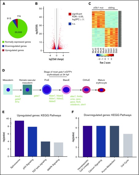
TGFβ- and Tp53-regulated genes are upregulated in sf3b1-mutant erythroid progenitors. (A) Representative chart of gene expression in gata1:eGFP+ cells from sf3b1 mutants. (B) Volcano plot displaying differentially expressed genes between gata1:eGFP+ cells from sf3b1 mutants and siblings. Significant differences are defined as FDR <0.05 and log2 fold change (FC) >1. Gray lines denote the fold-change threshold. (C) Heatmap of top 500 differentially expressed genes between gata1:eGFP+ cells from sf3b1 mutants and siblings. Each row is a gene and each column is a replicate. Mutant samples are shown on the left and sibling samples are shown on the right. (D) Schematic highlighting normally expressed hematopoietic genes in sf3b1-mutant erythrocytes and the stage at which they begin to be expressed (gene names in green). (E-F) Representative charts of significantly altered KEGG pathways in genes upregulated (E) or downregulated (F) in sf3b1-mutant erythroid progenitors compared with sibling controls as determined by mSigDB analysis. BasoE, basophilic erythroblast; OrthoE, orthochromatophilic erythroblast; ProE, proerythroblast.

