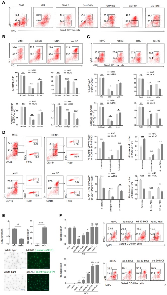
Figure 2.
Lnc-C/EBPβ promotes differentiation of PMN-MDSC but impedes Mo-MDSC in vitro. (A) Flow cytometry of inflammatory cytokine and tumor mediated CD11b+Ly6G+Ly6C− and CD11b+Ly6G−Ly6C+ MDSCs. 1D8, mouse ovarian carcinoma; 4T1, mouse breast cancer; B16, mouse melanoma. BMC, bone marrow cells; GM, GM-CSF. (B) Flow cytometry of Gr1+CD11b+, Gr1hiCD11b+, and Gr1lowCD11b+ MDSCs in lnc-C/EBPβ knockdown or exogenous lnc-C/EBPβ treated MDSCs. The percentages and absolute number of cells were compared (n = 3). (C) Flow cytometry of CD11b+Ly6G+Ly6C− and CD11b+Ly6G−Ly6C+ MDSC in lnc-C/EBPβ knockdown or exogenous lnc-C/EBPβ treated MDSCs. The percentages and absolute number of cells were compared (n = 3). (D) Flow cytometry of lnc-C/EBPβ nockdown and extinct lnc-C/EBPβ treated MDSCs. After culturing for 4 days, lnc-C/EBPβ knockdown and extinct lnc-C/EBPβ treated MDSCs were analyzed by staining using anti-Gr-1, anti-CD11b, and F4/80 antibody. The percentages and absolute number of cells were compared (n = 3). (E) QRT-PCR of mouse lnc-C/EBPβ in MDSCs after transfecting with lnc-C/EBPβ lentivirus (upper) and fluorescence microscopic imaging of green fluorescent protein in lentivirus infected cells (lower). (F) QRT-PCR of mouse lnc-C/EBPβ in cells transfected with different concentrations of lentivirus and flow cytometry of CD11b+ly6G+Ly6C−, CD11b+ly6G−Ly6C+ MDSCs in lnc-C/EBPβ lentivirus treated MDSCs. MOI, multiplicity of infection; kdLNC(kd), lentivirus/lnc-C/EBPβ shRNA; oeLNC(oe), lentivirus/lnc-C/EBPβ; kdNC and oeNC, control lentiviruses. Data are a representative of at least three experiments. Absolute number = total cell number × percentage of MDSC subsets. Two-tailed, paired T-test was used in B-F. *p < 0.05; **p < 0.05; ***p < 0.001. NS, no significant.

