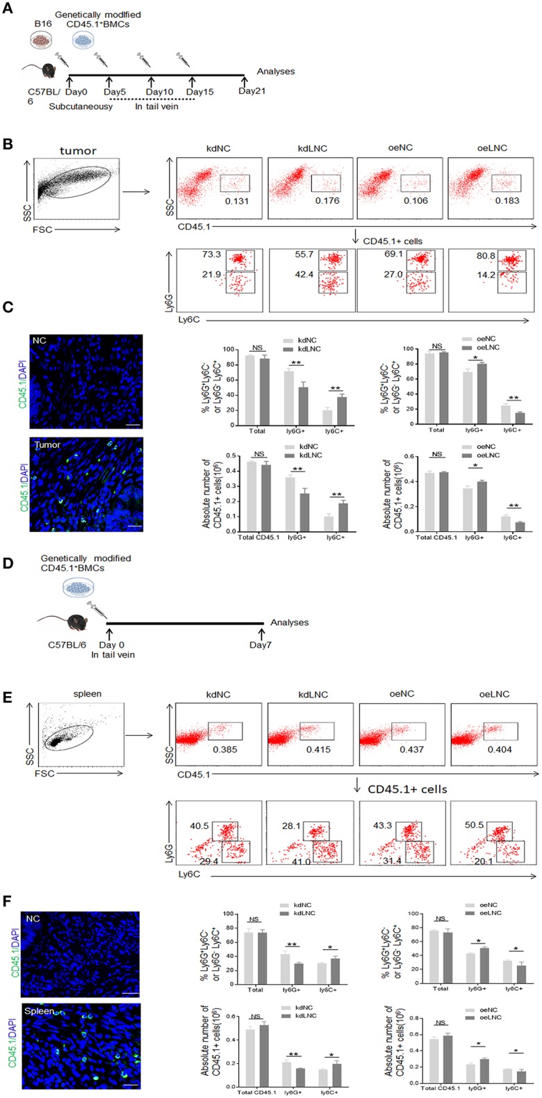
Figure 3.

Lnc-C/EBPβ promotes differentiation of PMN-MDSC but impedes Mo-MDSC in vivo. (A) Schematic of the experiment. Lnc-C/EBPβ lentivirus treated CD45.1+ BMCs were injected into B16 tumor bearing WT mice in tail vein, and after 21 days the tumor CD45.1 cells were analyzed. (B) Flow cytometry of MDSCs in mouse tumor after injecting lnc-C/EBPβ lentivirus treated CD45.1+BMCs. The percentage changes and absolute cell number of CD45.1+ cell of MDSC subsets were compared (n = 6). (C) Confocal microscopy of CD45.1+ cells in tumor. Green, CD45.1; blue, DAPI; NC, isotypic Ab. Scale bars = 100 μm. (D) Schematic of the experiment. lnc-C/EBPβ lentivirus treated CD45.1+ BMCs were injected into WT mice in tail vein, and after 1 week the spleen CD45.1 cell was analyzed. (E) Flow cytometry of MDSCs in mouse spleen after injecting lnc-C/EBPβ lentivirus treated CD45.1+MDSCs. The percentage changes and the absolute cell number of CD45.1+ MDSC subsets were compared (n = 6). (F) Confocal microscopy of CD45.1+ cells in spleen. Green, CD45.1; blue, DAPI; NC, isotypic Ab. Scale bars = 100 μm. kdLNC, lentivirus/lnc-C/EBPβ shRNA; oeLNC, lentivirus/lnc-C/EBPβ; kdNC and oeNC, control lentiviruses. Flow cytometry in (B,E) is a representative of at least three experiments. Absolute number = total CD45.1+ cell number in spleen or tumor × percentage of CD45.1+ MDSC subsets. Two-tailed, paired T-test was used in (B,D). *p < 0.05; **p < 0.05; NS, no significant.
