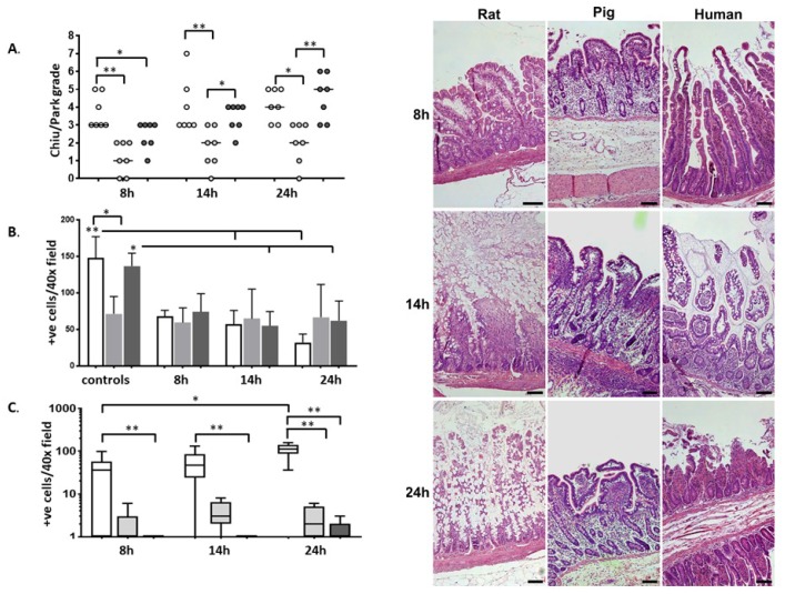
Figure 1.
Light microscopy of rat (white), pig (light grey), and human (dark grey) intestines after different periods of cold storage (CS). (A) Summary of the tissue injury (Chiu score) induced by CS with each dot representing one individual (n = 7) and the bar showing the median value; (B) goblet cell count; (C) enterocyte apoptosis quantified by caspase-3 positive cells (box plot showing the median, 5–95th percentile, and lowest and highest values at each time point). * p < 0.05, ** p < 0.01. A large number of apoptotic enterocytes (positive for active caspase-3) were found in rat intestines after 8 h of CS. Rat intestines had more apoptotic enterocytes than human intestines at all time points (Figure 1C). Right: representative microphotographs from each species at each of the three time-points (hematoxillin eosin stain, original magnification ×100, scale bar 100 microns).

