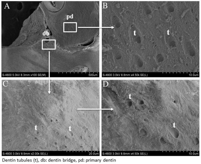
Figure 3.
Scanning electron micrograph after direct pulp capping using MTA. SEM analysis shows a well-mineralized tissue near the pulp exposure (A) and at higher magnification, many dentinal tubules through the reparative dentin bridge can be observed (C,D). The number of dentinal tubules is lower than that of the primary dentin (B).

