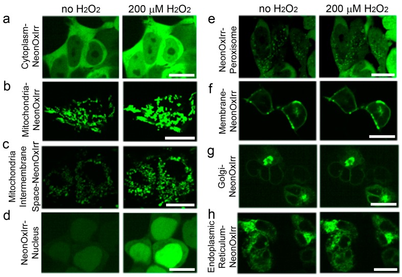
Figure 3.
Targeting and response of the NeonOxIrr indicator to external H2O2 in different compartments of live HEK293T mammalian cells. Confocal images of HEK293T cells transiently expressing the NeonOxIrr indicator in the cytoplasm (a), lumen of mitochondria (b), intermembrane space of mitochondria (c), nucleus (d), peroxisomes (e), plasma membrane (f), Golgi apparatus (g), and endoplasmic reticulum (h) are shown before and 5 min after H2O2 addition until a 200 µM final concentration. Scale bars – 20 µm.

