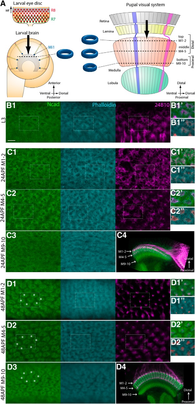
Figure 1.
N-cadherin and phalloidin visualize developing columns. A, Schematics of the developing larval and pupal visual systems. In the larval eye disc, R8 and R7 are sequentially differentiated behind the morphogenetic furrow. The NBs, located on the surface of the larval brain, produce Mi1 medulla neurons. In the pupal visual system, the retina, lamina, medulla, and lobula are topographically connected. Columns are identifiable along the planes indicated by dotted lines in the larval and pupal brains along the three layers (top, M1–M2; middle, M4–M5; bottom, M9–M10). B–D, The donut-like columnar patterns are identifiable in L3 (B1), 24 h APF (C1–C3), and 48 h APF stages (D1–D3) by Ncad (green), phalloidin (blue), and 24B10 (magenta). B1, Single layer of columnar pattern in L3 larval stage. C1–C3, D1–D3, Three layers of columnar patterns in 24 h APF (C1–C3) and 48 h APF (D1–D3) pupal stages. Layers M1–M2 (C1,D1), M4–M5 (C2,D2), and M9-M10 (C3,D3) are shown. Areas indicated by white rectangles are shown at right (B1,C1–C2,D1–D2). In contrast to the clear and regular patterns in layers M1–M2 and M4–M5 (C1,C2), the columnar pattern is irregular in layers M9-M10 at 24 h APF (C3), which become organized by 48 h APF (D3). Distal to the top, dorsal to the right. The following images are oriented in the same manner. C4, D4, Horizontal sections of 24 and 48 h APF brains to visualize the layers M1–M2, M4–M5, and M9-M10. Scale bar: B1, 5 μm.

