Figure 1.
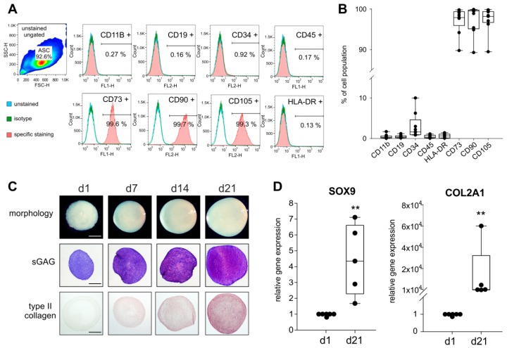
Analysis of mesenchymal stem cells (MSC)-specific surface markers and chondrogenic differentiation capacity of synovial adipose tissue-derived MSCs (sASC) obtained from osteoarthritis (OA) patients. (A) Human sASCs derived from OA patients were positive for CD73, CD90, and CD105 and were negative for CD11b, CD19, CD34, CD45 and human leukocyte antigen–DR isotype (HLA-DR) using FACS analysis (blue line—unstained negative control, green line—isotype control, red line—the target surface marker). (B) Quantification of MSC-specific marker expression on sASCs from eight different OA patients (n = 8). Data are presented as box plots with the 10th, 25th, 50th (median), 75th, and 90th percentiles. Each black circle represents an individual patient: (C) Macroscopic, histologic (sGAG) and immunohistochemical (type II collagen) analysis of untreated chondrogenic sASC pellets at day1, day 7, day 14, and day 21 of differentiation (bars, 500 μm). (D) Relative gene expression of SOX9 and type II collagen in untreated ASC pellets after 21 days of chondrogenesis. Data are presented as box plots with the 10th, 25th, 50th (median), 75th, and 90th percentiles. Each black circle represents an individual patient (n = 5–6). Significant p-values against untreated control are presented as ** = p < 0.01.

