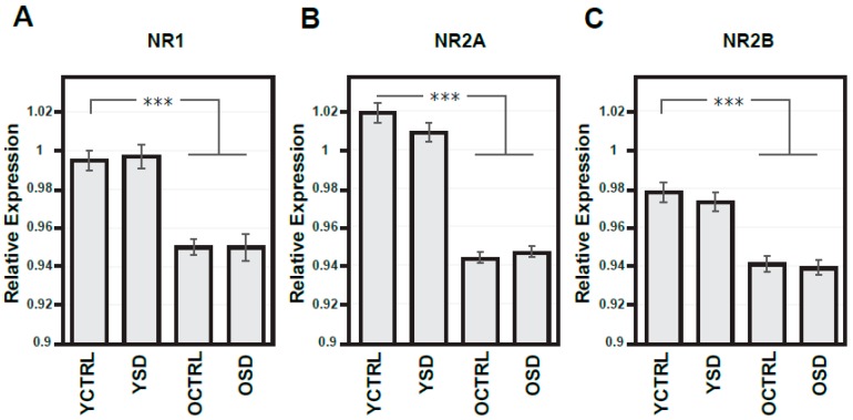
Figure 4.
Expressions of NMDA receptor subunits in the frontal cortex from both hemispheres in young adult and old rats exposed to acute sleep deprivation. Optical density of samples respective NMDA subunit bands (A–C) related to that of α-tubulin; results are presented as means ± SEM. Statistical significance (Student’s t-test) was calculated with respect to young adult controls (*** p < 0.001). SD—sleep deprivation, YCTRL—young adult controls (n = 12), YSD—young adult rats exposed to acute SD (n = 12), OCTRL—old controls (n = 12), OSD—old rats exposed to acute SD (n = 12).

