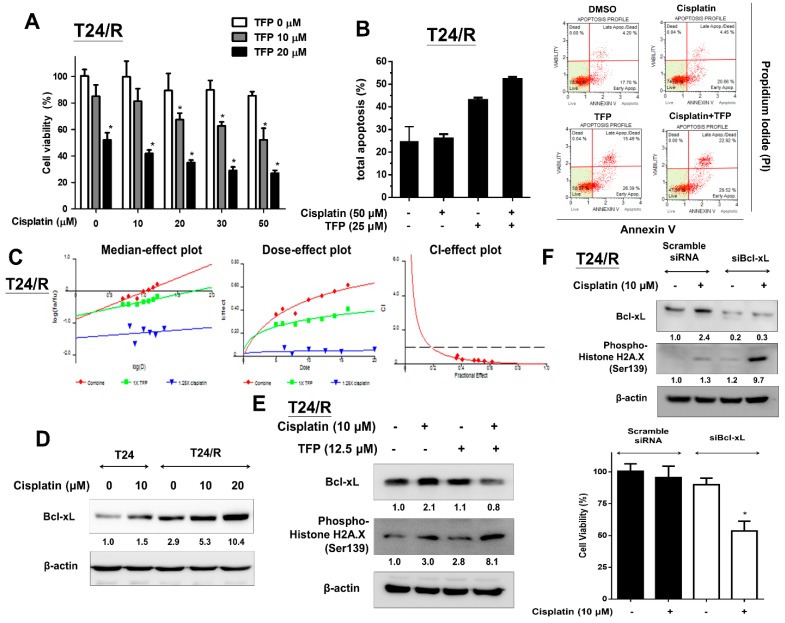
Figure 4.
TFP enhanced the antitumor effects of cisplatin on T24/R cells. The alleviation of cisplatin resistance was associated with concurrent suppression of Bcl-xL. (A) T24/R cells were treated with cisplatin (10–50 μM) or TFP (10 and 20 μM) alone or in combination for 24 h. Cell viability was determined using the MTT assay. (B) Cells were exposed to cisplatin (50 μM) or TFP (25 μM) alone or in combination for 24 h. Apoptotic cells were analyzed through FACS flow cytometry with propidium iodide and Annexin V-FITC staining. (C) T24/R cells were incubated in the presence of TFP, cisplatin, and in combination at the concentration ratio of 1:1.25 (TFP:cisplatin). Cell viability was measured by MTT assay after 24 h exposure. The median-effect plot, dose-effect plot and the combination index (CI)-effect plot for TFP, cisplatin, and the combination. The combination of TFP and cisplatin exhibited synergistic effects (combination index <1) in T24/R cells. (D) T24 parental cells and T24/R cells were treated with cisplatin (10 μM) and TFP (10–20 μM) separately. Cell lysates were collected and analyzed for Bcl-xL expression using Western blot analysis. (E) T24/R cells were treated with cisplatin (10 μM) or TFP (10–25 μM) alone or in combination for 24 h. Cell lysates were subjected to Western blot analysis of Bcl-xL and phospho-histone H2A.X (Ser 139). (F) T24/R cells were transfected with scrambled and Bcl-xL siRNA for 24 h, followed by cisplatin (10 μM) treatment for 24 h. Cell viability was determined using the MTT assay. Quantitative analyses of cell viability are presented as the means ± SD. The results shown are representative of at least three independent experiments. * p < 0.05 represents a significant difference between the indicated groups.

