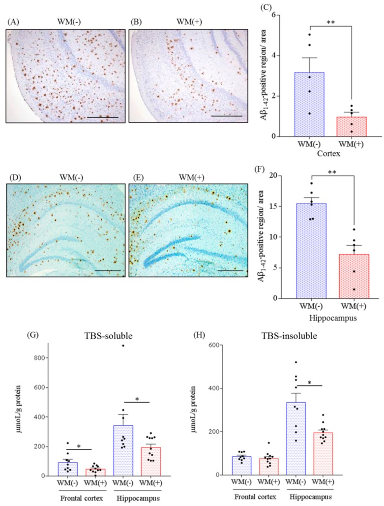
Figure 1.
Effects of WM peptide on β amyloid (Aβ) deposition in 5×FAD mice. For 3 months, 2.5-month-old transgenic 5×FAD and wild-type female mice were fed a diet either containing or not containing 0.05% w/w WM peptide. Percentage of Aβ1–42 positive area was detected by immunohistochemistry in the brains of the transgenic control mice [WM(−)] and transgenic mice fed on a diet containing WM peptide [WM(+)]. (A), (B), (D) and (E): The representative immunohistochemistry images including olfactory-entorhinal cortex and hippocampus for Aβ1–42 in transgenic mice with or without WM peptide, respectively. (C) and (F): Semiquantification for Aβ1–42 detected immunohistochemically in cortex and hippocampus, respectively. (G) and (H): The levels of Tris-buffered saline (TBS)-soluble Aβ1–42 and TBS-insoluble and TBS-T-soluble Aβ1–42, respectively, in the frontal cortex and hippocampus were measured by ELISA. Scale bars indicate 500 μm (A and B)and 400 μm (D and E), respectively. Data represent the mean ± SEM values. There were 5–7 mice in each group for immunohistochemistry and 9 control transgenic mice and 11 transgenic mice were fed on a diet containing the WM peptide for ELISA. The p-values shown in the graph were calculated via the Student’s t-test. * p < 0.05 and ** p < 0.01.

