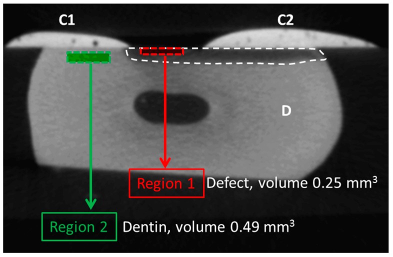
Figure 2.
Selection of the region of interest. In each sample, composite (C1) was used to cover part of the dentin defect, after which a defect (hatched, darkened area) was created using Buskes solution. Part of the defect was then covered by composite (C2), samples were scanned with a micro-CT for the first time, after which either control treatment or treatment with amine fluoride (AMF), enamel matrix derivatives (EMDs), or dentin matrix proteins (DMPs) was initiated. Following a second round of scanning and scan alignment, two regions of interests were created. Region 1 had a volume of 0.25 mm3 and was positioned within the top layer of the defect created by the Buskes solution. Region 2 had a volume of 0.49 mm3 and was selected as a background control, to rule out technical changes, e.g., due to the scanning procedure. The ratio of the two gray scale values of both regions was taken, i.e., (region 1/region 2) × 100%. This procedure was performed within the scans made before treatment and within the scans made after treatment (i.e., control (CON), AMF, EMP, and DMP groups) and subsequent incubation in artificial saliva.

