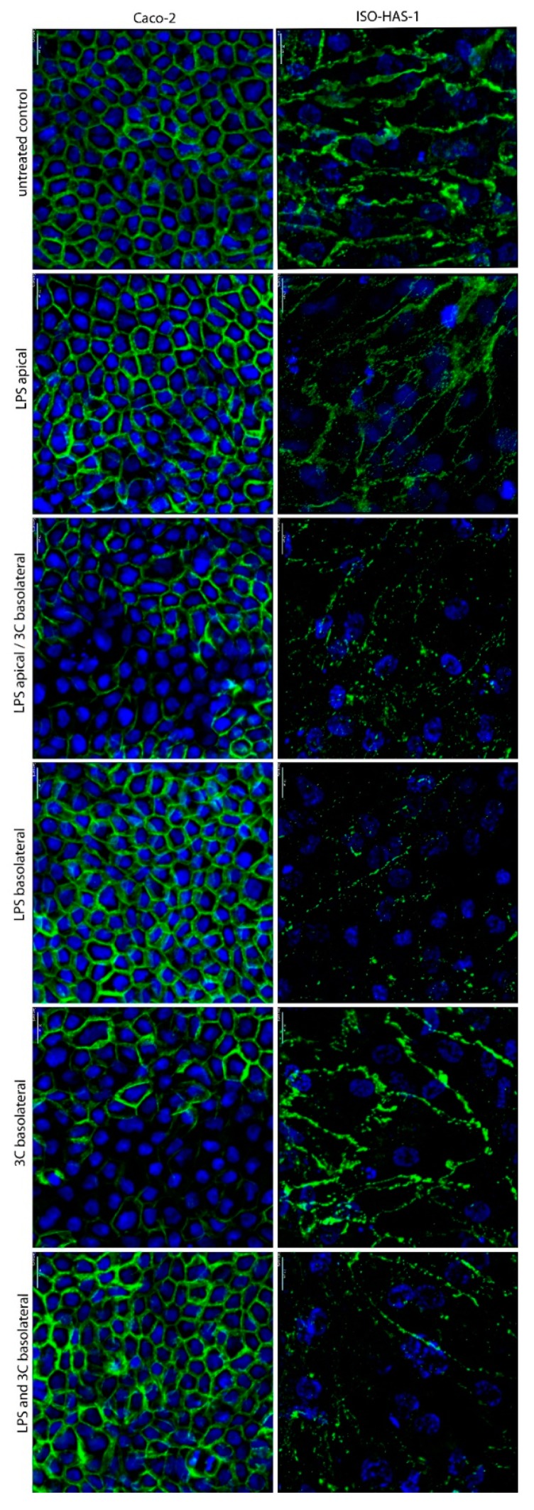
Figure 1.
Immunofluorescence staining of the coculture CC (Caco-2/ISO-HAS-1) after stimulation with different pro-inflammatory mediators for 48 h. apical: stimulation in the upper well (Caco-2), basolateral: stimulation in the lower well (ISO-HAS-1); LPS: lipopolysaccharide (1 µg/mL); 3C: cytokine mixture ((IL-1β (100 U/mL), TNF-α (600 U/mL) and IFN-γ (200 U/mL)); green signal: immunofluorescence of β-Catenin for both cell types Caco-2 (left column) and ISO-HAS-1 (right column); blue signal: nuclei stained with Hoechst 33342; scale bar: 20 µm.

