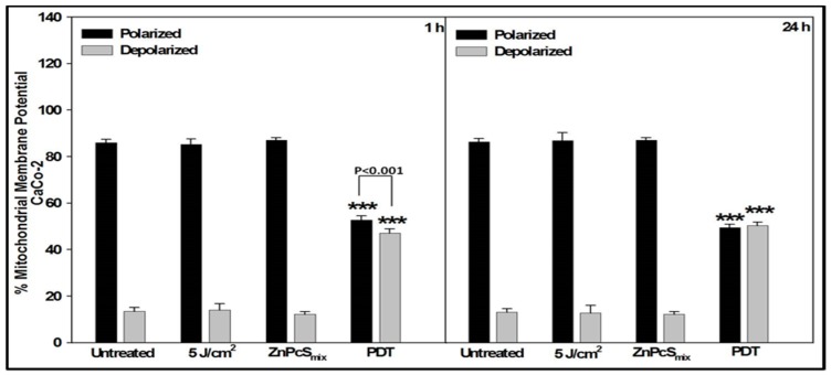
Figure 3.
Loss of mitochondrial membrane potential in Caco-2 cells was analyzed 1 or 24 h post-treatment by JC-1 staining using flow cytometry. Significant differences as compared to untreated cells as shown as *** p < 0.001. At both incubation periods, there was a significant loss of mitochondrial membrane potential in PDT treated cells.

