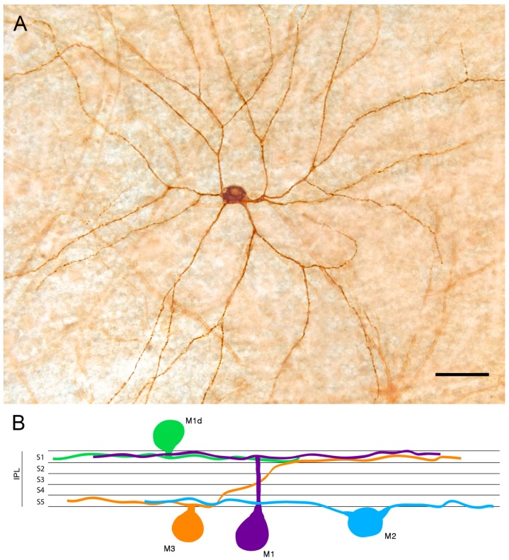
Figure 1.
Melanopsin-containing ganglion cells (mRGCs) detected by conventional immunostaining: (A) Immunostaining of a displaced M1 cell (M1d) mRGC found in wholemount human retinas. (B) Diagram showing the structure of the mRGC types depending on their soma location and dendrite stratification in the inner plexiform layer (IPL) S1 or S5. Scale bar: 50 μm. (Modified from Ortuño-Lizaran et al., 2018) [42].

