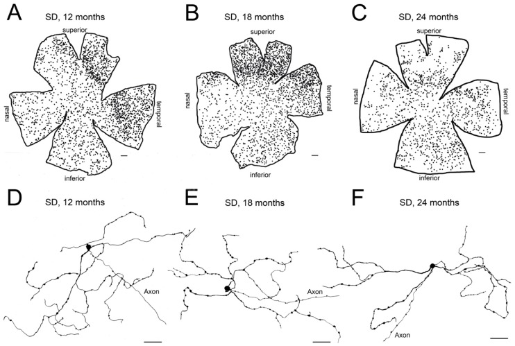
Figure 3.
Age-related changes of melanopsin ganglion cells in control Sprague–Dawley rat retinas: (A–C) Representative drawings of wholemount retinas from Sprague–Dawley (SD) rats at 12 (A), 18 (B), and 24 (C) months-of-age. (D–F) Representative drawings of the soma and complete dendritic field of mRGCs from a region of the central retina (between the superior and nasal quadrants) of Sprague–Dawley rats at 12 (D), 18 (E), and 24 (F) months-of-age. Drawings were made using a camera lucida and reveal immunostained mRGCs. Scale bar: A–C, 500 μm; D–F, 50 μm. (Modified from Lax et al., 2016) [63].

