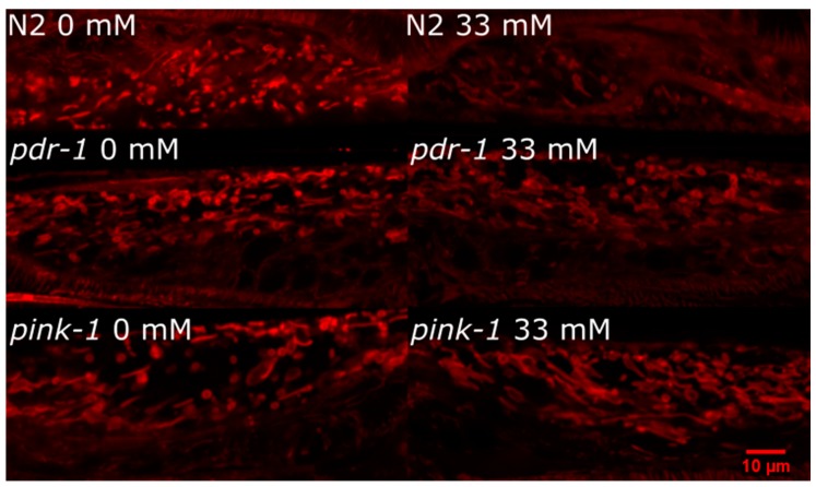
Figure 7.
Mitochondrial morphology is not dramatically altered in pink-1 or pdr-1 mutants after 6-OHDA exposure compared to N2 (wild-type) animals. For experiments, worms (wild-type and mitophagy-deficient mutants crossed into the BY200 transgenic reporter strain) were exposed to 6-OHDA as in other experiments. Following exposure, mitochondrial morphology was visualized using MitoTracker CMXRos (incubated with worms for final 4 h of 24 h recovery after exposure). Confocal imaging was performed on animals immobilized with tetramisole and mounted on an agarose pad as described in Methods.

