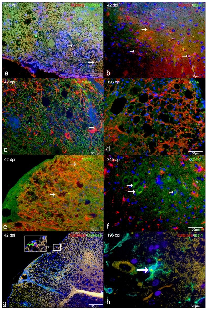
Figure 8.
Immunofluorescence in TMEV-DL lesions: (a) 8OHdG and CNPase as well as oligodendrocytes positive for 8OHdG (double labeling, yellow, arrow) in the white matter of spinal cord of TMEV-DL, (b) 8OHdG and Iba1, showing that macrophages/microglia are positive for 8OHdG (double labeling, yellow, arrow) in TMEV-DL, (c) MDA and GFAP double labeling (yellow, arrow) in TMEV-DL, (d) MDA and GFAP, showing that astrocytes do not significant co-localize with MDA (double labeling, yellow) in TMEV-DL, (e) SOD2and GFAP as well as astrocytes positive for SOD2 (double labeling yellow, arrow) in TMEV-DL, (f) SOD2and GFAP double labeling (yellow) in TMEV-DL, some SOD2 positive cells share characteristics of neurons (arrow), (g) Catalase and CNPase, showing that oligodendrocytes can be positive for catalase (double labeling, yellow, green arrow within the insert) in TMEV-DL, (h) Catalase and Iba1 as well as macrophages/microglia positive for catalase (double labeling, yellow, arrow) in TMEV-DL.

