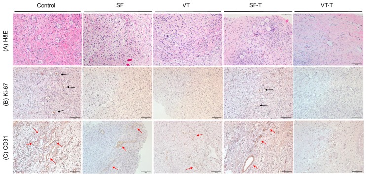
Figure 3.
Histopathological findings of the human ovarian tissues after cryopreservation and xenotransplantation. (A) Hematoxylin and eosin staining (H&E) of human ovarian tissues in the five groups. (B) Histological features of human ovarian tissues in the five groups evaluated with immunohistochemistry staining for Ki-67, a marker of cell proliferation. Black arrows indicate the cells stained with Ki-67. (C) Histological features of human ovarian tissues in the five groups evaluated with immunohistochemistry staining for CD31, a marker of angiogenesis. Red arrows indicate the cells stained with CD31. Original magnification ×200. Abbreviations: SF, cryopreserved with slow freezing technique and thawed group; VT, cryopreserved with vitrification technique and thawed group; SF-T, the group xenotransplanted after slow freezing and thawing; VT-T, the group xenotransplanted after vitrification and thawing.

