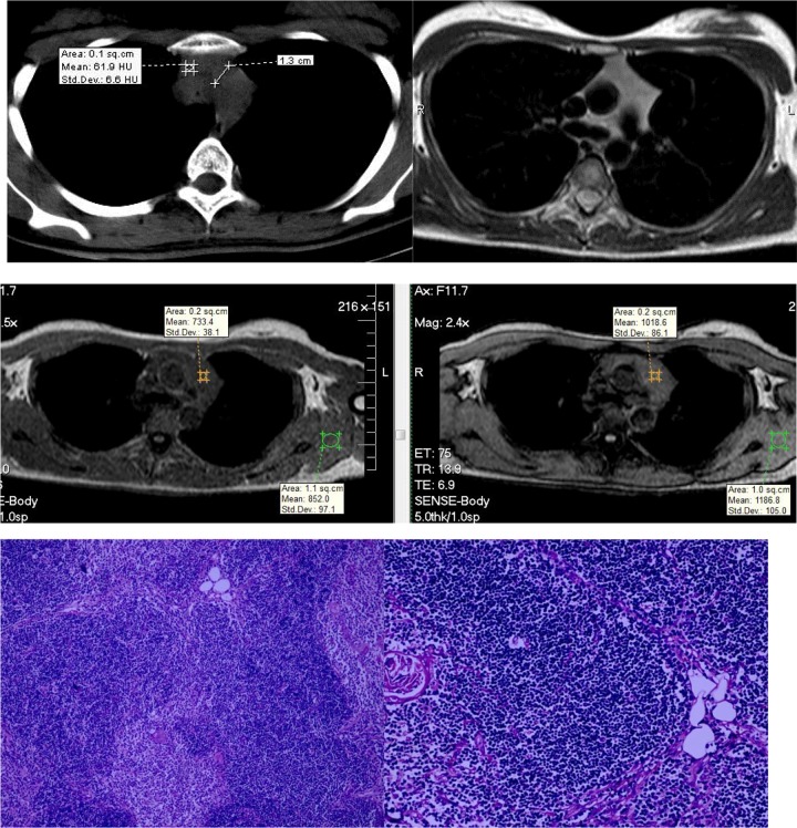
Figure 4.
A 22-year-old woman with TLH. Both CT and MRI showed a bilobed shaped soft tissue homogeneous gland with straight margins without the signal intensity loss in the opposed-phase image relative to the in-phase image. The CSR value was 1.0. A histopathologic image exhibits preserved architecture of the thymus, consisting of cortex, medulla, and Hassall corpuscles with lymphoid follicles and minimally fatty infiltrated thymic tissue. CSR indicates chemical shift ratio; CT, computed tomography; MRI, magnetic resonance imaging; TLH, thymic lymphoid hyperplasia.

