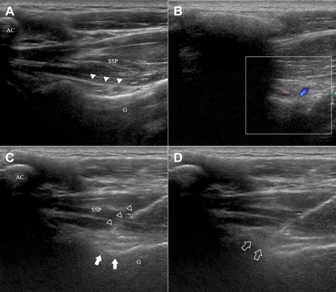
Figure 1.
Ultrasonographic images of the suprascapular nerve block (SSNB) at the spinoglenoid notch. (A) An ultrasonographic image of the suprascapular nerve. The hyperechoic line (solid arrowheads) is the inferior fascia of the supraspinatus (SSP). The suprascapular artery and nerve are located below this line. (B, C) After locating the suprascapular artery with a Doppler scan, the needle (open arrowheads) is inserted using a medial in-plane approach such that the tip is placed close to the spinoglenoid notch (solid arrows), where the suprascapular neurovascular bundle is located. As the bundle is barely visible, particular caution should be taken when performing this procedure. AC, acromion; G, glenoid. (D) Local anesthetic spreading around the suprascapular nerve after the injection (open arrows).

