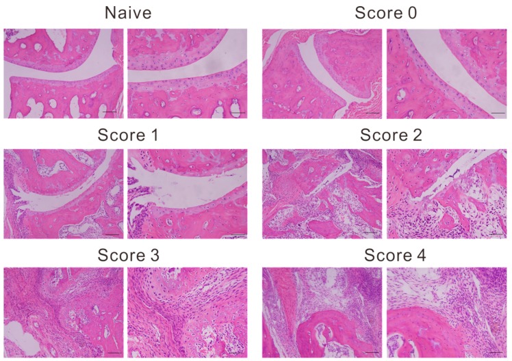
Figure 4.
Representative images of histopathology sections. Naive and arthritic joint from each group, stained with hematoxylin and eosin (left panel magnification, ×100; right panel magnification, ×200). Photomicrographs of left hind ankles from five representative mice are shown for each clinical score group.

