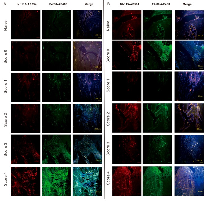
Figure 5.
Confocal microscopy demonstrates that Vsig4+ F4/80+ macrophages increased according to the severity of arthritis. Immunofluorescence microscopy of CIA joints left hind ankles having different clinical scores. DBA-1 mice were immunized with type II collagen in complete Freund’s adjuvant. Slides were incubated with Nb119 labelled by AF549 (red) and AF488-labelled anti-F4/80 (green). Cell nuclei were stained with 4′,6-diamino-2-phenylindole (DAPI) dye (blue), colocalization of F4/80 and Vsig4 were shown in the third panel (white). (A) Scale bar = 200 μm. (B) Scale bar = 400 μm. Photomicrographs of left hind ankles from representative 5 mice are shown for each clinical score group.

