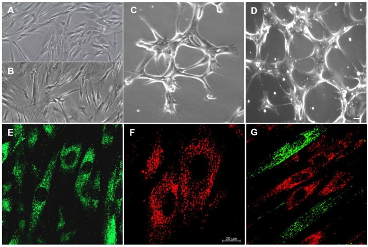
Figure 2.
Morphological analyses. (A) Undifferentiated hPDLSCs observed at light microscopy. (B) E-hPDLSCs observed at light microscopy. (C,D) E-hPDLSCs cultured on Cultrex® observed at light microscopy. (E) Undifferentiated hPDLSCs evaluated under CLSM (PKH67, green fluorescence). (F) E-hPDLSCs evaluated under at CLSM (PKH26, red fluorescence). (G) Coculture of hPDLSCs and E-hPDLSCs. Scale bar: 20 µm. Magnification: 10× (A–D). Magnification: 20× (E–G).

