Figure 3.
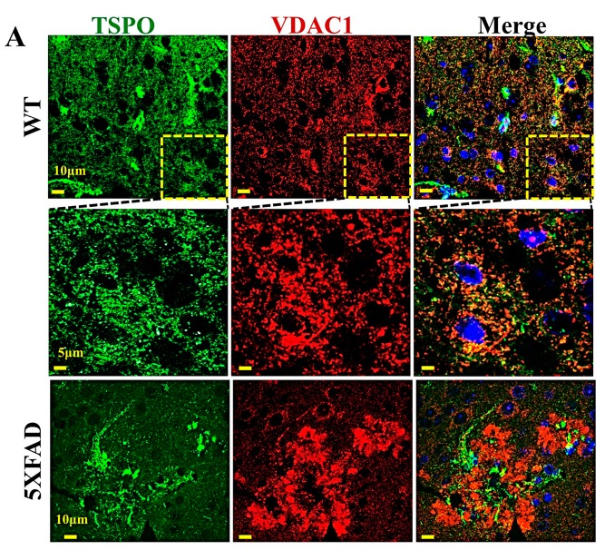
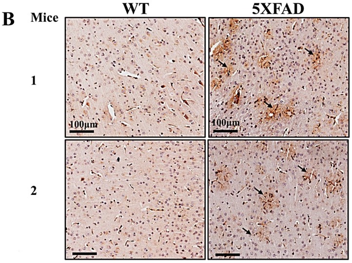
TSPO and VDAC1 are over-expressed in the brains of transgenic mice. (A) Cross-sections of brains from wild-type (WT) and 5XFAD transgenic mice, immunofluorescently stained for TSPO or VDAC1. Formalin-fixed and paraffin-embedded 5 μm-thick brain sections were deparaffinized, rehydrated, and subjected to antigen retrieval in 0.01 M citrate buffer (pH 6.0). For confocal fluorescence microscopic imaging of immuno-stained brain sections from WT and 5XFAD transgenic mice, the tissues were stained with anti-TSPO or anti-VDAC1 antibodies. Nuclei were stained by DAPI. Immunofluorescent staining were performed using mouse anti-VDAC1 (1:1000) and rabbit anti-TSPO (1:500) antibodies, followed by incubation (2 h, 25 °C) with secondary ant-rabbit Alexa-flur-488 or anti-mouse Alexa-Flu 555 (1:1000) antibodies. The cells were then stained with DAPI and viewed with an Olympus IX81 confocal microscope. (B) For immunohistochemistry, endogenous peroxidase activity was blocked by incubating the sections in 3% H2O2 for 15 min, after which the slides were washed and incubated overnight at 4 °C with primary rabbit anti-TSPO antibodies (1:200) and then for 2 h with anti-rabbit (1:500) secondary antibodies conjugated to horseradish peroxidase (HRP). Sections were washed and incubated with the HRP substrate, DAB. Images were collected at 20× magnification using a microscope (Leica DM2500). Non-specific control experiments were conducted using the same protocols but omitting incubation with primary antibodies. Arrows points to β plaques enriched with TSPO-expressing microglia.


