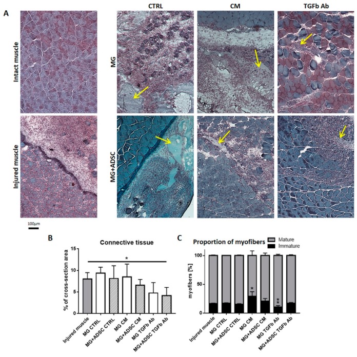
Figure 4.

Analysis of skeletal muscle and connective tissue morphology. (A) Morphology of skeletal muscles (blue) stained with Harris hematoxylin and Gomori Trichrome dye, at 7 day of regeneration. Intact muscles, injured muscles, and muscles which received Matrigel or Matrigel with ADSC pretreated with control (CTRL), myoblast-conditioned (CM), or supplemented with antibody against TGFβ (TGFb Ab) medium. Arrows indicates localization of Matrigel. (B) Area occupied by connective tissue analyzed on cross-sections of injured muscles and muscles which received Matrigel or Matrigel with ADSC pretreated with control (CTRL), myoblast-conditioned (CM) or supplemented with antibody against TGFβ (TGFb Ab) medium. (C) Analysis of the proportion of mature and immature muscle fibers present in regenerating skeletal muscles of all analyzed groups. For each experimental group n ≥ 3. Data are presented as mean ± SD. * represent results of Student’s t-test: * p ≤ 0.05, ** p ≤ 0.01. Bar - 100 µm.
