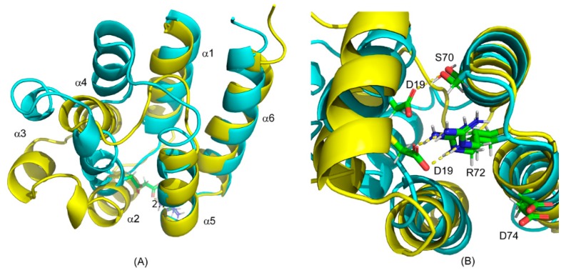
Figure 3.
Superposition of CS-Rosetta structural models of the free and ERK2-bound forms of PEA-15. (A) Superimposition of the free form (yellow) with the bound-form models (cyan). The ERK2-bound PEA-15 displays minimum changes in helices α1, α5, and α6, but significant conformational change in helices α2, α3, and α4. (B) Positions of charge-triad residues in the free (yellow) and ERK2-bound models (cyan). The D19 residue moves significantly, while the R72 and D74 residues mostly stay at their original positions. The hydrogen-bonding interactions between the D19 and R72 residues in the free-form structure is replaced by the D19–S70 hydrogen bond in the ERK2-bound structure.

