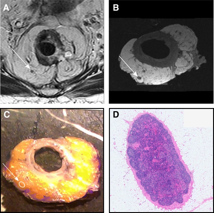
Fig. 2.
An exemple of the matching process. Anatomically matched lymph node, measuring 2.5 mm in short axis, seen in a transaxial T2 weighted sequence perpendicular to the tumour, b transaxial T1-weighted sequence MRI of the surgical specimen, c in the finding-by-finding description using the photographed slices arrayed numerically, and d at microscopy, using hematoxylin & eosin stain at 1.5x magnification, where no malignant growth was seen

