Fig. 7.
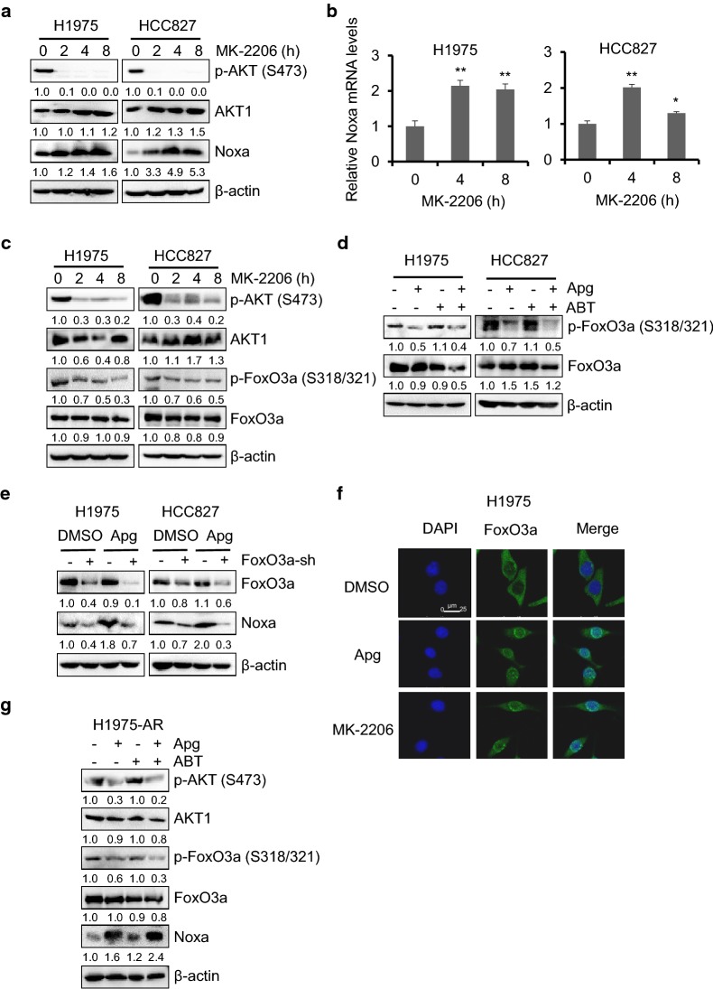
Dephosphorylation of FoxO3a by apigenin upregulated Noxa expression in EGFRm tumor cells. a H1975 and HCC827 cells were treated with MK-2206 (1 µM) for up to 8 h. Cells were harvested and the expression levels of AKT and Noxa were detected by the immunoblotting. b H1975 and HCC827 cells were treated with 1 µM MK-2206 for up to 8 h. Then the total RNA was extracted and the Noxa mRNA level was quantified by real-time PCR. The data represents mean ± SD (n = 3). *p < 0.05, and **p < 0.01. c H1975 and HCC827 cells were treated as described in a, total and phosphorylated AKT and FoxO3a was examined by Western blotting. d H1975 and HCC827 cells were treated with Apg (15 µM), ABT (2 µM) alone or comb for 1 day. Cells were harvested, and the expression levels of FoxO3a were detected by the immunoblotting. e H1975 and HCC827 cells infected with the ctrl-sh or FoxO3a-sh lentivirus were treated with 15 µM Apg. Apoptotic death rates were analyzed, and expression of FoxO3a and Noxa were analyzed by Western blotting. f Immunofluorescence staining of FoxO3a in H1975 cells. Cells were treated with apigenin or MK-2206 for 8 h. Cells were then stained for FoxO3a and nucleus using Alexa Fluor 488-conjugated antibody (green) and DAPI (blue), respectively. Representative staining cells are shown. g H1975-AR cells were treated with Apg (15 µM) and ABT (2 µM), alone or in combination for 1 day. Cells were harvested, and the expression levels of p-AKT, AKT, p-FoxO3a, FoxO3a and Noxa were examined by Western blotting

