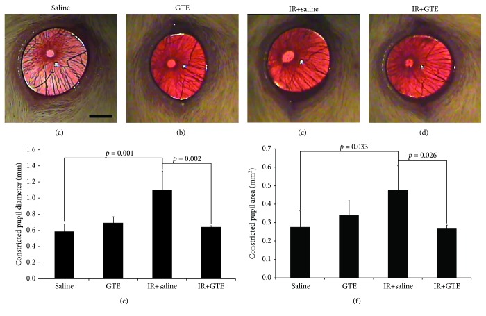
Figure 3.
Pupillary light reflex analysis for the green tea extract treatment effect on normal and ischemia-injured rats. Retinal ganglion cell (RGC) function was evaluated by the pupillary light reflex on day 14 after ischemic reperfusion (IR) injury. Pupillary light reflex was measured by the pupil constriction. (a) Surviving RGCs in the saline-fed normal rats. (b) Surviving RGCs in the green tea extract- (GTE-) fed normal rats. (c) Surviving RGCs in the saline-fed rats with ischemic injury. (d) Surviving RGCs in the GTE-fed rats with ischemic injury. (e) Quantitative analysis of the constricted pupil diameter. (f) Quantitative analysis of the constricted pupil area. Data was presented as mean ± standard deviation. Scale bar: 2 mm.

