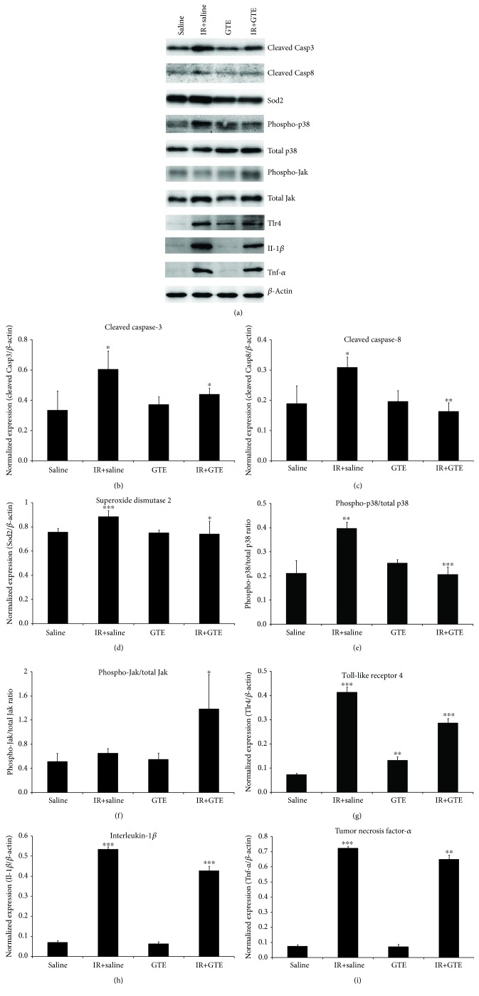
Figure 4.
The expression analysis of cell apoptosis, oxidative stress, and inflammation-related proteins in the retina of ischemia-injured rats with green tea extract treatment. (a) Protein expression was evaluated by the immunoblotting analysis on day 2 after ischemic reperfusion injury. Relative fold change analyses on (b) caspase-8 (Casp8), (c) toll-like receptor 4 (TLR4), (d) interleukin-1β (Il-1β), (e) tumor necrosis factor-α (TNF-α), (f) phospho-p38 (p-p38), and (g) phospho-Jak (p-Jak). β-Actin was used as housekeeping protein for normalization. Data was presented as mean ± standard deviation. Compared to the saline-fed normal rats: ∗∗p < 0.01, ∗∗∗p < 0.001. Compared to the saline-fed ischemia-injured rats: #p < 0.05, ##p < 0.01, ###p < 0.001.

