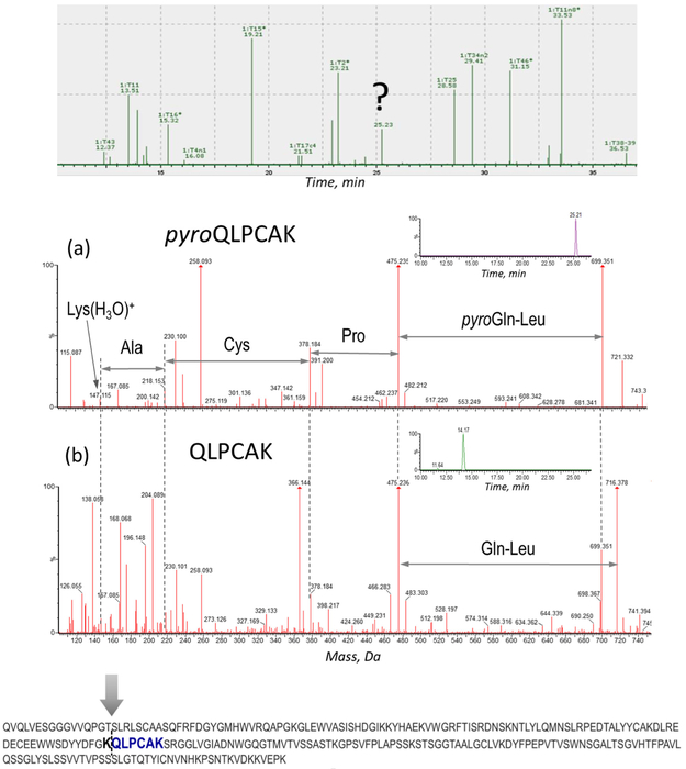
Figure 7.
Identification of the clipping location based on MSE de novo sequencing of the unassigned tryptic component. Top: unassigned peak on the chromatogram. Middle: assignment of pyroQLPCAK (a) and its non-modified analog QLPCAK (b); XIC of the corresponding peptides: 699.3 Da (pyroQLPCAK) (a, inset), 716.3 Da (QLPCAK) (b, inset), (0.1 Da extracted mass window). Bottom: Fab portion of CAP256 heavy chain showing the clipping site and the identified tryptic peptide marked in bold

