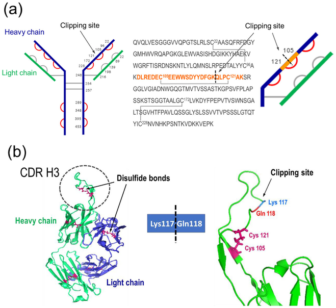
Figure 8.
Clipping site identification. (a) A protruding loop in the CDR-H3 region (left); clipping location with a disulfide bond preventing the non-reduced bNAb to fall apart into two fragments (right). (b Disulfide bond arrangement, planar view: Cys residues are numbered, verified disulfide bonds marked in red. The sequence of the heavy chain Fab region is shown, along with the corresponding schematic view zoomed into the clipping site: the tryptic semidigested peptide (highlighted in orange) identified as a peptide dimer (tryptic component held together by a disulfide bond), covering the clipping site area

