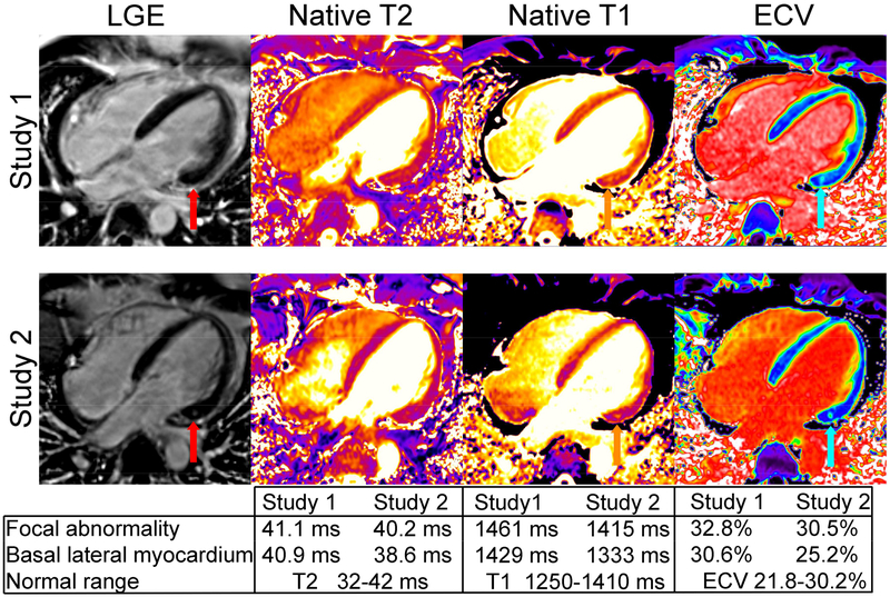
Figure 1. Cardiac MRI studies were performed following recovery from severe Ebola virus disease on clinical illness day 32 (study 1) and 11 weeks later (study 2).
Study 1 showed mildly reduced ejection fraction (LVEF 52%). Late gadolinium enhancement (LGE) was initially unremarkable (red arrow, study 1). There was evidence of mild edema or inflammation of the midwall of the basal inferolateral segment of the left ventricle with a focal abnormal spot on native T1 and ECV (orange and cyan arrows) but also milder abnormalities in surrounding myocardium excluding the spot (see table). Quantitative maps of myocardial T2 showed a non-significant elevation in mid-myocardial T2 in the interventricular septum. On study 2, the T1 and ECV abnormalities normalized except for a sub-gram region localized to the region of most severe abnormality seen on the initial study. Interval development of fibrosis was evident on LGE images (red arrow on study 2). LVEF remained mildly abnormal (51%).

