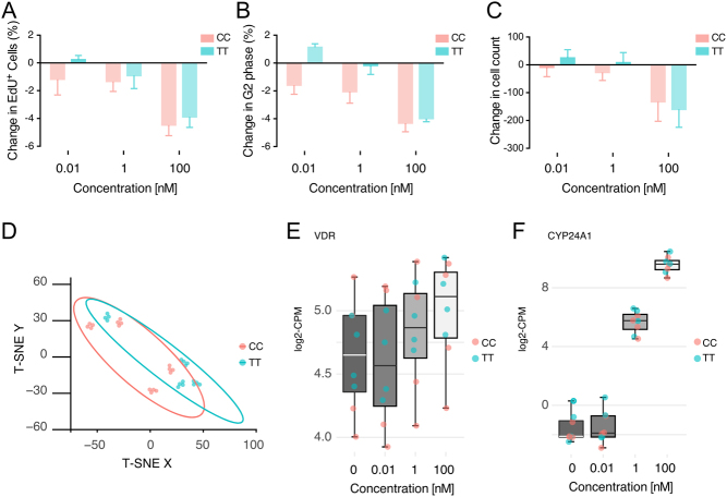
Figure 1.
Using high-throughput imaging analysis, primary human muscle cells with the Taq1 polymorphism were stained and imaged for DAPI and EdU incorporation after 48-h exposure to DMSO alone or 0.01 nM, 1 nM and 100 nM 1,25(OH)2D3. Data are presented for change in total cell count (A), change in the percentage incorporation of EdU-positive cells (B) and change in the percentage of cells that are G2 positive (C), as the mean ± s.e.m. TT versus CC. (D) Unsupervised clustering using tSNE of gene expression of all cells and all used dosages of vitamin. (E and F) VDR and CYP24A1 expression following various dosages of vitamin D.

 This work is licensed under a