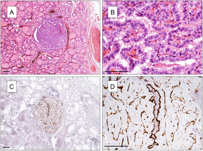
Figure 2.
NOTCH1 expression in a papillary microcarcinoma and in the normal surrounding thyroid tissue. (A and B) H&E staining (in B microcarcinoma at higher power view). (C and D) NOTCH1 staining. NOTCH1 positivity is restricted to microcarcinoma (in the center; in D at higher power view), while normal thyroid tissue is negative. (A, B and C) Magnification 40×, (B, C and D) 100×. Scale bar 100 µm.

 This work is licensed under a