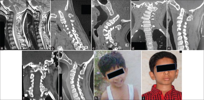
Figure 2.
Images of a 6-year-old boy. (a) T2-weighted magnetic resonance imaging shows basilar invagination, assimilation of atlas, and C2–C3 fusion. (b) Sagittal computed tomography scan in flexion shows basilar invagination, assimilation of atlas, and C2–C3 fusion. (c) Sagittal computed tomography in extension shows no significant reduction in basilar invagination. (d) Preoperative coronal computed tomography showing the deformity and torticollis. (e) Postoperative computed tomography showing reduction of basilar invagination and craniovertebral realignment. (f) Postoperative coronal computed tomography shows intra-articular implant on the right side and plate and screw fixation on the left side. (g) Delayed postoperative computed tomography scan shows craniovertebral fusion. (h) Preoperative clinical image. (i) Postoperative image showing reduction of torticollis

