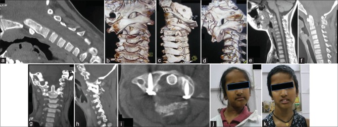
Figure 3.
Images of a 9-year-old female. (a) Computed tomography scan shows marked basilar invagination. (b) Three-dimensional computed tomography scan shows complete rotatory atlantoaxial dislocation. (c) Posterior view showing the rotatory dislocation. (d) Oblique view of three-dimensional computed tomography scan shows the rotatory abnormality. (e) T1-weighted magnetic resonance imaging shows basilar invagination. (f) Postoperative computed tomography shows reduction of basilar invagination. (g) Coronal computed tomography scan shows reduction of the facetal malalignment. (h) Computed tomography showing the implant and the facets in alignment. (i) Axial computed tomography scan shows screws in the facets of atlas. (j) Preoperative clinical image of the patient. (k) Postoperative image showing reduction of the torticollis

