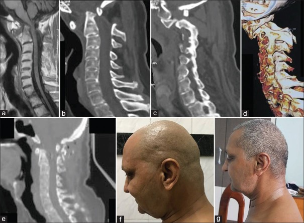
Figure 6.
Images of a 51-year-old male patient. (a) T1-weighted magnetic resonance imaging showing the craniovertebral junction and atlantoaxial instability. (b) Computed tomography scan showing the atlantoaxial dislocation. (c) Computed tomography scan of the facets showing Type 1 atlantoaxial facetal instability. (d) Three-dimensional computed tomography scan shows anterior translation of both facets of atlas over facets of axis. (e) Postoperative computed tomography scan shows craniovertebral fixation. (f) Preoperative clinical picture shows fixed flexion deformity of the head. (g) Postoperative clinical image shows normalization of head posture

