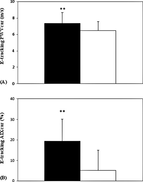
Figure 2.

(A) Comparison of local (carotid) pulse wave velocity (PWVcar) in patients with verified coronary artery disease (CAD) with age‐ and sex‐matched apparently healthy control subjects. (B) Comparison of local (carotid) augmentation index (AIxcar) in patients with verified CAD with age and sex‐matched apparently healthy control subjects. These measurements were carried out with Doppler echo‐tracking method. Data are presented as mean ± standard deviation.  = CAD group (n = 35).
  = control group (n = 35). **
P < 0.01.
