
Figure 1.
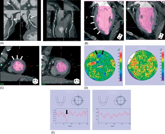
Representative case of myocardial perfusion defect. (A) A 78‐year‐old man presented with effort angina. 64‐multidetector computed tomography showed severe coronary artery stenosis in the proximal portion of the left anterior descending coronary artery (arrow, left panel). (B) and (C) Myocardial perfusion imaging demonstrated subendocardial hypoenhancement in the anterior wall of the left ventricle (arrow, left panel). (D) Myocardial perfusion map showed the clear hypoenhancement area in the anterior region (arrow, left panel). (E) Histograms of myocardial perfusion showed decreased perfusion in the anterior wall (arrow, left panel). After coronary artery bypass surgery this myocardial hypoenhancement improved (B–D, right panel).