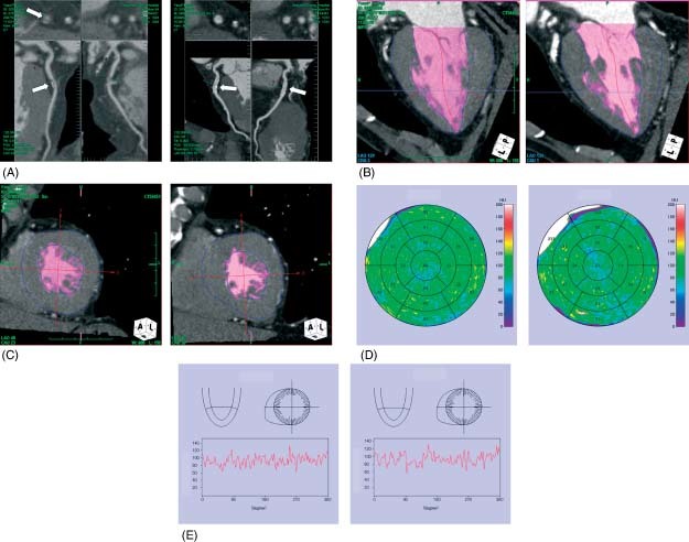
Figure 2.

Representative case of normal myocardial perfusion. (A) A 74‐year‐old woman with chest pain underwent MDCT, which showed significant coronary artery stenosis in the proximal portion of the left anterior descending coronary artery (arrow, left panel). (B) and (C) Myocardial perfusion imaging demonstrated no hypoenhancement area in the left ventricle (left panel). (D) and (E) Myocardial perfusion map and histograms of myocardial perfusion showed no hypoenhancement area in the left ventricle (left panel). After coronary stent implantation (A; arrow, right panel) there is no hypoenhancement area in the left ventricle (B–D; right panel).
