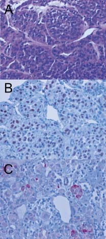
Figure 4.

Histology of the resected tumor tissue from the left ventricle. The tumor consisted of large, polygonal tumor cells (A) arranged in an alveolar pattern and variably sized compact cell nests surrounded by well‐vascularized and partially hyalinized fibrous stroma. Mitotic figures were scarce. Several tumor cells contained brightly eosinophilic intracytoplasmic crystalloid inclusions. Immunohistochemistry (B) revealed strong nuclear reactivity for TFE3. In addition, isolated tumor cells (C) were also reactive for desmin. Abbreviations: TFE3, transcription factor binding to IGHM enhancer 3.
