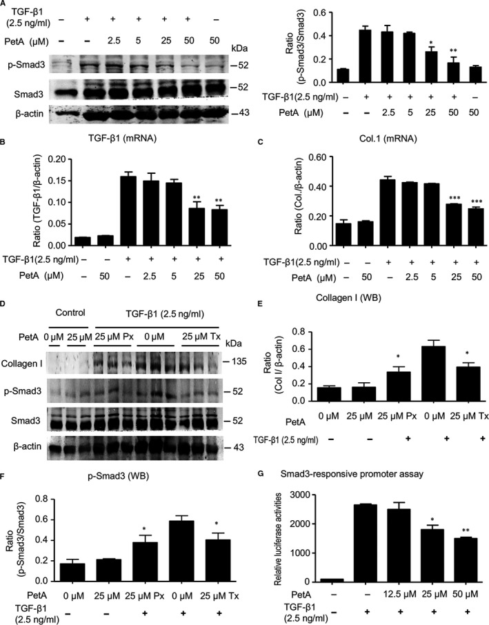
Figure 6.

Petchiether A (PetA) attenuates collagen I expression by inhibiting transforming growth factor‐β1 (TGF‐β1)‐induced phosphorylation of Smad3 in cultured HK‐2 cells. A, HK‐2 cells were pre‐treated with the indicated dose of petA (2.5, 5, 25, 50 μmol/L) or vehicle for 12 h before stimulation with TGF‐β1 (2.5 ng/mL) for another 12 h and examined for Smad3 phosphorylation by Western blotting. Representative Western blots and quantitative analysis of Smad3 phosphorylation are shown. TGF‐β1 mRNA expression (B) and (C) collagen 1 mRNA expression were measured using quantitative real‐time PCR. D, HK‐2 cells were pre‐treated with petA (25 μmol/L) or vehicle for 12 h before (Px group) or after (Tx group) stimulation with TGF‐β1 (2.5 ng/mL) for another 12 h. Representative Western blots show that the addition of petA (25 μmol/L) can inhibit TGF‐β1‐induced up‐regulation of collagen I expression (E) and Smad3 phosphorylation (F). G, PetA inhibits Smad3‐specific luciferase reporter p(CAGA)‐luc in HK‐2 cells. HK‐2 cells were cotransfected with the p(CAGA)‐luc plasmid, followed by pre‐treatment with the indicated dose of petA (12.5, 25, 50 μmol/L) or vehicle for 4 h before stimulation with TGF‐β1 (2.5 ng/mL) for another 12 h. Luciferase activity in the control group was set at 100%. Compared with the increased p(CAGA)‐luc reporter activity in petA‐untreated HK‐2 cells under TGF‐β1 stimulation, petA at a dose of 25 μmol/L significantly inhibited (CAGA) reporter activity. Data represent the mean ± SEM for at least three independent experiments. *P < 0.05, **P < 0.01 compared with petA‐untreated cells under TGF‐β1 stimulation. PetA Px, PetA prevention; PetA Tx, PetA treatment
