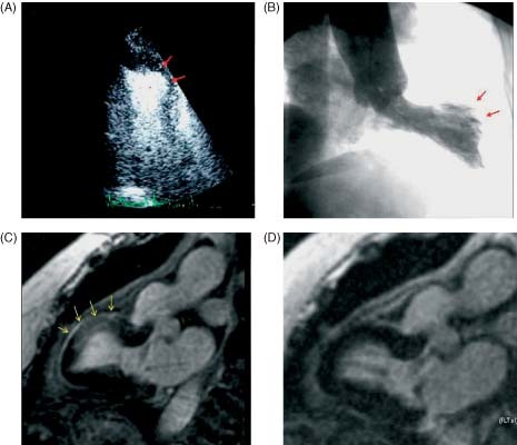
Figure 2.

Patient with transient midventricular dyskinesis. (A) Contrast echocardiogram (systolic image), apical 2‐chamber view showing dyskinesis in the middle segment of the anterior wall (red arrows). (B) Angiographic ventriculography, showing similar findings to those described by echo (red arrows). (C) CMR, sequence of 2‐chamber LGE showing hyperintensity in the area of abnormal segmental wall motion (middle segment of the anterior wall) (arrows). (D) Complete normalization after 30 days. Abbreviations: CMR, cardiac magnetic resonance; LGE, late gadolinium enhancement.
