FIG 5.
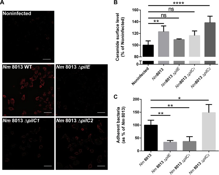
Ceramide release and CRP formation in response to isogenic ΔpilC1 or ΔpilC2 meningococcal mutants. (A) HBMEC were grown to confluence in 8-well Ibidi μ-slides and were infected with wild-type (WT) strain N. meningitidis 8013 or isogenic meningococcal mutant N. meningitidis 8013 ΔpilE, N. meningitidis 8013 ΔpilC1, or N. meningitidis 8013 ΔpilC2 for 4 h or were left noninfected. Cells were washed, fixed in FA, and stained with an anticeramide antibody and secondary Cy5-conjugated goat anti-mouse IgM (red). Images were captured using a Nikon Eclipse Ti-E inverted microscope with a 20× objective lens. Bars, 100 μm. The results of one of three reproducible experiments are shown. (B) HBMEC were infected with wild-type strain N. meningitidis 8013 or isogenic mutant N. meningitidis 8013 ΔpilE, N. meningitidis 8013 ΔpilC1, or N. meningitidis 8013 ΔpilC2 or were left noninfected for 4 h. Surface ceramide levels were determined by flow cytometry, and data are represented as the relative levels of ceramides on the host cell surface. (C) HBMEC were infected with the N. meningitidis 8013 wild-type strain or the indicated mutants (the ΔpilE, ΔpilC1, or ΔpilC2 mutant) for 4 h at an MOI of 100. Adhesion was determined by a gentamicin protection assay. Error bars represent the mean ± SD. One-way ANOVA with Dunnett’s post hoc test was used to determine significance. *, P < 0.05; **, P < 0.01; ****, P < 0.0001; ns, not significant.

