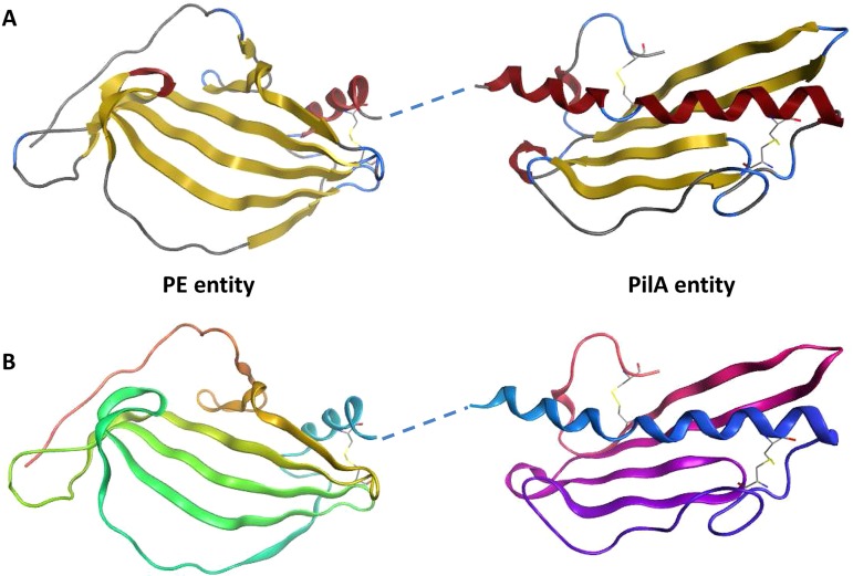
FIG 8.
Crystal structure of PE-PilA. (A) The β-strands are shown in yellow, and the α-helices are shown in red. The linker region was not resolved but is shown here as a dashed line linking the two entities. (B) The amino acid succession is shown from orange in the PE entity (N terminus) to pink in the PilA entity (C terminus).

