FIG 8.
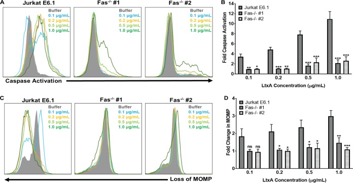
Fas mediates caspase activation and mitochondrial membrane permeabilization in response to LtxA. (A) Jurkat E6.1 cells and Jurkat Fas−/− clones were treated with various concentrations of LtxA for 24 h. Caspase activation was assessed using a fluorescent polycaspase reagent and flow cytometry. Increases in fluorescent signal are directly related to the amount of active caspases within a cell. Representative images are shown. (B) Cells were gated on buffer-treated controls, and fold change in caspase activation was determined using FlowJo software. (C) Jurkat E6.1 cells and Jurkat Fas−/− clones were treated with various concentrations of LtxA for 24 h, and mitochondrial membrane permeability was assessed via MitoTracker staining and flow cytometry. Decreases in fluorescent signal are associated with compromised mitochondrial membranes. Representative images are shown. MOMP, mitochondrial outer membrane protein. (D) Cells were gated on buffer controls, and fold change in mitochondrial membrane permeabilization was determined using FlowJo software. Cell populations are indicated according to the legend on the figure. Data represent the average of at least three independent experiments. Error bars represent SEM. The significance of differences between results for Jurkat E6.1 cells and those for each Jurkat Fas−/− clone was determined using Student's t test. *, P ≤ 0.05; **, P ≤ 0.01; ***, P ≤ 0.001; ns, not significant.

