Figure 1.

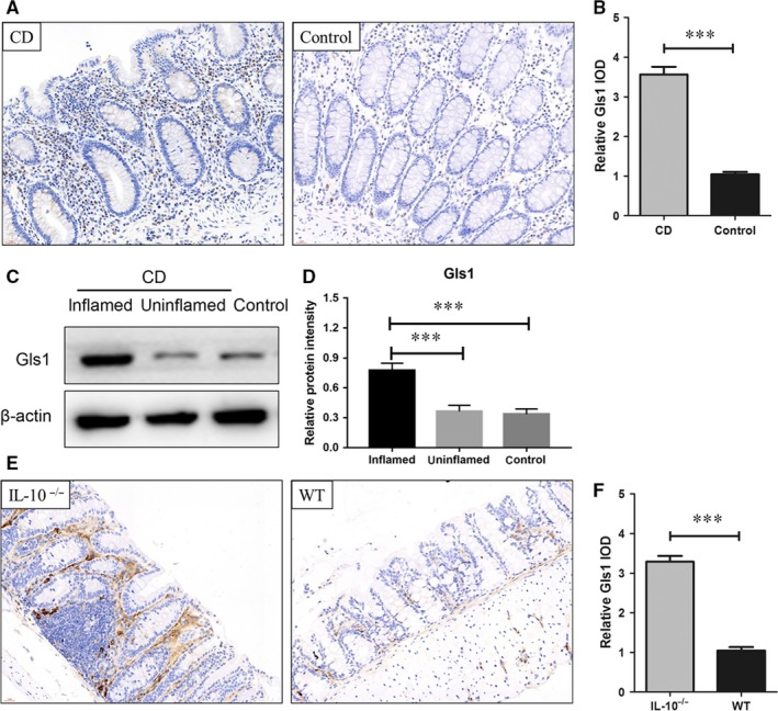
Gls 1 is highly expressed in the intestines of CD patients and Il‐10 –/– mice. Immunohistochemical staining with an antibody that recognizes Gls1 was performed on the intestines of control and CD patients (A). The quantitative analysis presented in (B) shows Gls1 expression in the intestines of CD (n = 13) and control patients (n = 17). (C,D) Western blot analysis of Gls1 in the intestinal mucosa in the intestines of CD patients (inflamed and uninflamed areas) and control patients. (E,F) The expression of Gls1 in the intestines of Il‐10–/– mice and WT mice (n = 8 in each group). CD, Crohn's disease; Gls1, glutaminase 1; IOD, integrated optical density; WT, wild‐type. The data are presented as the relative IOD ± SD. ***P < 0.001
