
Figure 6.
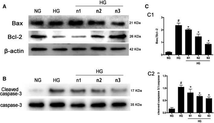
Apoptosis level reduced after nicorandil treatment in high glucose‐induced H9c2 cardiomyocyte. A: Western blot analysis of bax and bcl‐2 in high glucose‐induced H9c2 cardiomyocyte after nicorandil treatment with different concentrations for 24 h. B: Western bolt analysis of cleaved caspase‐3 in high glucose‐induced H9c2 cardiomyocyte after nicorandil treatment. HG (33.3 mmol/L), NG (5.5 mmol/L), n1: nicorandil (10 µmol); n2: nicorandil (50 µmol); n3: nicorandil (100 µmol). # P < 0.05 compared with NG; *P < 0.05 compared with HG, Data are means ± SD